

Services
Diagnostic Service
Radiology Imaging
New PET-CT Centre
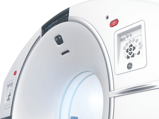
PET-CT Centre of Evangel Hospital is equipped with GE Healthcare Discovery MI positron emission tomography/computer tomography (Digital PET-CT). It is currently the most advanced PET- CT imaging technology and also a future trend. Comparing to traditional analogue system in the market, digital PET-CT has a higher sensitivity and image resolution. The new digital system could significantly reduce examination time and radiopharmaceutical dose up to 50%, allowing decrease absorption of radiation dose by examinees as compared to traditional systems. The digital detectors also could improve accuracy and help to identify small and early lesions.
What medical information could be gathered from PET-CT?
- Distinguishing cancer at early stage
- Identifying location and structure of tumour
- Grading and staging of cancer
- Determining condition of metastasis, if any
- Assisting in treatment planning
- Assessing treatment effectiveness
- Monitoring cancer recurrence if any
Advantages of New PET-CT Scanner DISCOVERY MI*
- Significant Reduction of Scanning Time & Radiation Dose
Digital PET detector converts received radiation information directly to image signal. It increases the sensitivity of the equipment, allowing scanning time and radiation dose to be reduced. - ASiR-V Reconstruction Technology
Reducing image noise and enhancing image quality. - Up to 2x Improvement of Image Quality & Accuracy
Innovative reconstruction technology including the combination of Time-of-Flight (TOF) and Q.Clear, which improves image quality and accuracy . - Increasing the Detectability of Small Lesions
Improves the visualization of small lesions so that early cancer/abnormalities can be easily detected.
*Depends on patient condition and examination items

MRI
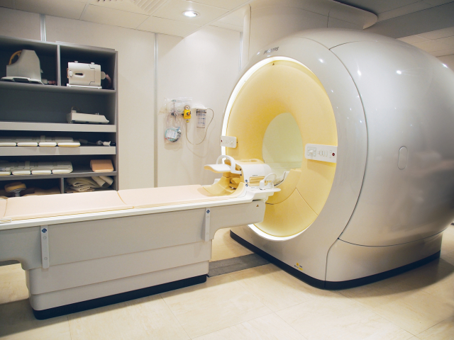
Evangel Hospital is equipped with high technology digital Magnetic Resonance Imaging (MRI) scanning system and is operated by professional radiologist and radiographers for the provision of high-quality imaging service and diagnosis. The high-quality images provided by our Philips Ingenia 1.5T, which is a fully digital, high resolution premium MR can improve diagnostic accuracy and hence the provision of the best treatment plan.
Magnetic Resonance Imaging (MRI) is a high-end medical imaging technology that uses a powerful magnetic field, radio waves, and combined with precise computer technology to generate detailed multiplanar images of your body.
Scope of Services:
- Cerebral vascular disease and cerebral tumor
- Artery disease (including angioma, atherosclerosis causing partial or total occlusion of blood vessels and others)s
- Heart disease (including ischemic heart disease, myocardial disease, congenital heart disease and others)
- Peripheral vascular disease
- Skeletal joint and soft tissue disease
- Vertebral column and sports injury
- Kidney and kidney vascular disease
- Cancer
- Hepatobiliary disease
- Gynecologic and breast disease
Advantages of MRI:
- Non-invasive and does not use ionizing radiation
- Particularly useful for the scanning and detection of abnormalities in soft tissue structures in the body, such as ligaments and cartilage, soft organs such as the brain, heart and eyes.
- Can provide information about how the blood moves through certain organs and blood vessels, allowing problems with blood circulation, such as blockages, to be identified.
- Possess high ability to differentiate tissue structures. The current accuracy of MRI is generally superior to both CT scan and ultrasound scanning
Computed Tomography (CT) Scanning
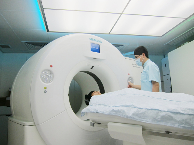
Evangel Hospital is equipped with the SIEMENS Dual Source CT Somatom Definition Flash. It provides faster speed, higher quality images and with lower radiation dose when compared to the conventional single source CT scanner.
A computerized tomography (CT) scan uses sophisticated x-ray technology and combines a series of X-ray images taken from different angles around your body and uses computer technology to create cross-sectional images (slices) of the bones, blood vessels and soft tissues. CT scan images provide more-detailed information than plain X-rays do.
Scope of Services:
- Coronary scanning
- Brain scanning
- Thorax scanning
- Abdomen scanning
- Pelvis scanning
- 3D Angiogram
- Carotid Angiogram
- Circle of Willis Angiogram
- Renal Arteries
- Pulmonary Angiogram
- Peripheral Angiogram
The clinical benefits of Dual Source CT Scanner:
- The fast scan time of the scanner allows the scanning of patients with irregular heart rates, atrial fibrillations and increased heart rate due to nervousness without the use of medicine to slow down the heart rate, like β-Blocker in a cardiac scan. It reduces the risk of medicinal induced allergy and yet obtains clear and accurate coronary images.
- The fast scanner can cover a wider imaging range in a short period of time, so the time required for the contrast medium to stay in the blood vessel or related organs can be reduced, resulting in a significant reduction in the total injected dose. Reducing the amount of contrast reduces the risk of contrast -induced nephropathy.
- Breath-hold technique is no longer required to maintain motion-free (clear) images for this fast scanner. It is particularly beneficial to elderly and children who are difficult to hold the breath for a long time. It reduces recall for examination due to suboptimal quality images.
- The radiation dose may be reduced up to 60% when compared with the conventional CT scanner.
Digital Mammography
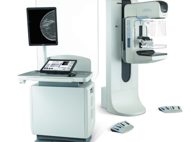
Evangel Hospital is equipped with advanced HOLOGIC Selenia Dimensions 3D Mammography System that provides superb image quality, high productivity and couples with a variety of advance clinical application software. Its helps to provide appropriate and efficient imaging service to the patients. Besides, its Genius 3D Mammography can detect early breast cancer and reduce false positive recalls.
Advantages of ESWL
- 41% increase in detection of invasive cancer VS 2D mammogram.
- A 3.7-second tomosynthesis scan time regardless of breast thickness.
- Low patient dose as fewer exposures are required.
- Clinically superior exams with comparable radiation dose to conventional 2D mammogram.
- Reduced recalls due to false positives by up to 40%.
- More comfortable by less the compression time.
DEXA Bone Densitometry
本院配備GE Lunar Prodigy雙能量骨質密度檢查儀,能夠進行全身體質密度及身體成分的一次性掃描,掃描範圍更廣。雙能量X光骨質密度檢查(DEXA)是一種評估特定部位骨頭中鈣質和礦物質數量的方法,它使用兩種不同穿透力的X光能量來進行。DEXA的放射劑量比傳統肺部X光低,具有相對安全和準確的特點。
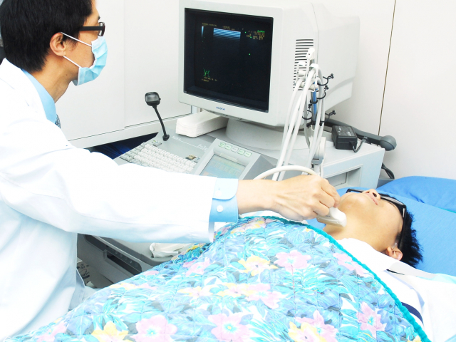
Ultrasound Scanning
Ultrasound imaging (sonography) uses high-frequency sound waves to capture the body’s internal organs in real time, through the use of a transducer (probe) and high technology imaging technique.
In an ultrasound examination, a thin layer of gel is applied to the skin so that the ultrasound waves are transmitted from the transducer through the gel into the body.
Unlike X-ray imaging, there is no ionizing radiation exposure associated with ultrasound imaging and it can visualize soft tissues that x-ray cannot.
Advantages of Ultrasound Scanning
- Non-radiation, non-invasive examination
- It is comparatively fast, convenience and comfortable
Scope of Service
- Obstetric and fetal scanning: to diagnose the normal growth of fetus
- Color Doppler scanning: to diagnose obstruction of peripheral blood vessels
- Ultrasound guided core tissue biopsy of tumor, e.g. breast cancer
- Thyroid Gland
- Male and female genitals
- Pelvic scanning
- Breast scanning

HA Referral Discount
Evangel Hospital cooperates with the HA hospitals, provide discount offer to referrals by HA, you are welcome to contact us for details of the offer.