

Services
Allied Health Service
Optometric Service
After completing a detailed optometry examination, you will receive an eye examination report. Many people often don't know what tests were performed or how to read this report. Below are some guidelines to better understand your eye examination report.
|
Refraction 屈光檢查 |
RE 右眼 |
LE 左眼 |
|
Sphere 近/遠視 |
-1.00 | +0.50 |
|
Cylinder 散光 |
-0.50 | -0.75 |
|
Axis 散光軸 |
180 | 180 |
|
Prism 稄鏡 |
||
|
Dist. VA 遠視力 |
1.0 | 1.0 |
|
ADD 老花 |
+2.00 | +2.00 |
|
Near VA 近視力 |
1.0 | 1.0 |
*FULL RX NOT EYEGLASSES PRESCRIPTION
| Color Vision色覺檢查 |
Normal 正常 |
|
| Binocularity 雙眼協調效能 | Normal 正常 | |
| Stereopsis立體感 | Normal 正常 | |
| Amsler chart 阿姆斯勒方格表 |
RE 右眼 | Normal 正常 |
| LE 左眼 | Normal 正常 | |
| Ocular Health眼睛健康 | ||
| RE 右眼 | External外部 | Normal 正常 |
| Internal 內部 | Normal 正常 | |
| LE 左眼 | External外部 | Normal 正常 |
| Internal 內部 | Normal 正常 | |
| Intra Ocular Pressure 眼內壓 | ||
| RE 右眼 | 18.5 mmHg | Normal 正常 |
| LE 左眼 | 17.8 mmHg | Normal 正常 |
| Suggestion 建議 |
Annual eye examination 周年眼睛檢查 |
- Refraction:
This refers to the examination of the eyes for conditions such as myopia, hyperopia, and astigmatism.
Sphere (Myopia): Represented with a “-” sign, e.g., -2.00 indicates 200 degrees of myopia.
Sphere (Hyperopia): Represented with a “+” sign, e.g., +2.00 indicates 200 degrees of hyperopia.
Cylinder: Represented with a “-” sign, e.g., -2.00 indicates 200 degrees of astigmatism. (In some older notations, a “+” sign was used, but this is rare now.)
Axis: The shape of an astigmatic eye resembles a rugby ball, and the astigmatic axis is used to indicate the angle of the longer axis. It is generally not of concern, but is necessary for the lens manufacturer when making glasses.
ADD: Represented with a “+” sign, e.g., +2.00 indicates 200 degrees of presbyopia.
Visual Acuity - VA (Dist. VA & Near VA): Used to indicate the ability to see clearly, with several representations, such as: "Decimal": 0.1, 0.5, 1.0 / "Snellen": 6/60, 6/12, 6/6 (commonly used). 1.0 and 6/6 represent the same visual acuity, which is considered standard vision. The smaller the decimal value or the larger the Snellen denominator, the worse the vision, i.e., 0.8 or 6/8 is clearer than 0.2 or 6/30. - Color Vision:
This is to check for color blindness or color deficiency. Complete color blindness is rare, with red-green color deficiency being more common. - Binocularity:
Various methods are used to check if both eyes can coordinate properly. This primarily involves checking for strabismus, which can be further classified as “manifest strabismus” and “latent strabismus.” Optometrist will decide whether follow-up or referral is necessary based on the condition. - Stereopsis:
Primarily used to aid in assessing binocular coordination efficiency. Stereopsis is generally poorer in individuals with poor binocular coordination or significant differences in vision between the two eyes. - Amsler Chart:
One method to test for macular degeneration. If the grid appears deformed, blurry, or missing, it may indicate a problem with the macula. - Ocular Health:
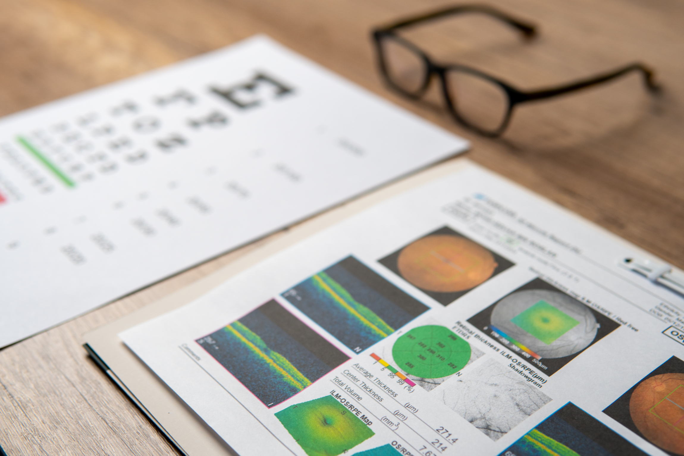
Various instruments such as fundus photography and slit lamps are used to examine eye structures like the retina, lens, and cornea for issues such as cataracts and diabetic retinopathy. - Intra Ocular Pressure:
Non-contact tonometers measure intraocular pressure. Combined with fundus examination, they help assess the risk of glaucoma. Normal intraocular pressure ranges from 8-22 mmHg.
Optometrist will make appropriate recommendations based on the examination results, such as referrals to ophthalmologists or other professionals, or regular follow-ups.